Hydra Data Pre-Processing Issue

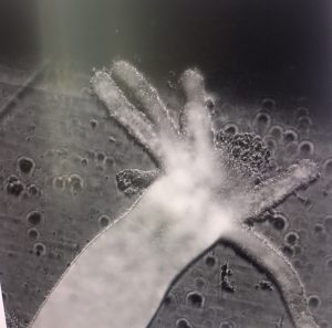
After fixing the issue of keeping the hydras from being flattened by the cover slip, we began collecting low-resolution images of the hydras to start running algorithms for reconstructing a high-resolution image.

We chose a sample data set with one stack of images and ran the Generatescript.py code. By executing the sh file that it created, we ran into a problem in which no high resolution image was generated. To see if the problem lay with the program or the images, we tried running the code with a flatworm data set imaged last summer. This succeeded in producing the high resolution image, confirming that the issue relates to our hydra data somehow.

Currently we suspect that the one stack of data is not sufficient for the pre-processing procedure, so we are currently trying with a five-stack hydra data set for the GenerateScript.py. Once the program stops running, we will see if this is in fact the source of the problem or not.

Data Collection of Hydras

Now that all the computers are set up except for the one with the monitor that will be using for data processing, we started collecting  low resolution image stacks from the hydras we received from Swarthmore’s biology department.

By placing the hydra into a dish and stabilizing it with a square plastic cover, we are able to observe the hydra under the Fourier Ptychographic microscope and collect photos using the camera.

We took photos with three different diameters of the LED matrix, specifically 9, 11 and 13. By lighting up different ranges of LED lights, we were able to create three low resolution data sets with 1 low resolution stack per diameter. After taking these datasets, we will decided that the 10x magnification is best if we want to be able to image the entire hydra rather than a part of that hydra.

Shift-add algorithms and the iterative reconstruction algorithm will be implemented once the computer with the algorithms is set up.

Notes on “Efficient illumination angle self-calibration in Fourier ptychography” by R. Eckert, Z.F. Phillips,L.Waller

The paper focuses on illumination angle self-calibration for Fourier ptychographic microscopy (FPM).  However, there appears to be errors in FPM due to shifts or rotations of the LED array, source instabilities, nonplanar illuminator arrangements, or sample-induced aberrations. The paper proposes  a two-pronged-angle self-calibration method that uses both preprocessing (brightfield calibration) and iterative joint estimation (spectral correlation calibration) that is quicker and more robust to system changes than state- of-the-art calibration methods.

For the Brightfield calibration, the following algorithm is used to perform the task.

For Spectral Correlation Calibration, we can see from the figure below that cost function is lower when both BC and SC are implemented.

The paper have presented a novel two-part calibration method for recovering the illumination angles of a computational illumi- nation system for Fourier ptychography. We have demon- strated how this self-calibrating method makes Fourier ptychographic microscopes more robust to system changes and sample-induced aberrations. The method also makes it possible to use high-angle illuminators, such as the quasi-dome, and nonrigid illuminators, such as laser-based systems, to their full potential.


References: 

R. Eckert, Z.F. Phillips,L.Waller.”Efficient illumination angle self-calibration in Fourier ptychography”, 2018 Optical Society of America

Notes on “Deep Learned Optical Multiplexing for Multi-Focal Plane Microscopy”

As was addressed in the last paper about FPM, it takes a long time and usually costly to scan a specimen and produce a high quality picture. This can be solved by reconstructing 69 photos taken under different LED pattern and using deep learning, the microscope can be trained to be able to produce a high-quality picture by taking one best photo, which shortens the time by a factor of 69.

However, such methods only satisfy situations where the sample given is thin enough so that the camera only needs to focus once. When encountering with circumstances where the sample is thick, the picture taken can be blurred due to the different distance each layer is from the camera. To improve the microscope’s functionality, digital refocusing is required during the process of reconstructing and training.

With assumptions of geometric optics, the 3-dimensional is assumed to modulate only the intensity instead of the phase. Instead of adding all the photos taken from which the obtained image would simply be that when all LED lights are on, the LED stack was sheared and resulting images were then added together. The shift-add algorithm would produce an example as shown below.

After completing the shift-add algorithm, the microscope is trained by using deep learning to achieve the goal of producing high-resolution picture in one shot of the photo, increasing the speed of scanning. Deep learning is divided up into training and evaluation and the algorithm is shown below.

Using this type of training and fine-tuning, the experiment is able to produce a result as shown in the graph.

References: 

Yi Fei Cheng, Ziad Sabry, Megan Strachan, Skyler Cornell, Jake Chanenson, Eva-Maria Collins, and Vidya Ganapati. “Deep Learned Optical Multiplexing for Multi-Focal Plane Microscopy” Department of Engineering and Biology, Swarthmore College

Equipment purchase sugguestions

LED Dome

No response has yet been received from Spectral Coded Illumination, Inc and the inquiry e-form is questionably functional.

DiffuserCam

DiffuserCam consists of a raspberry pi camera and tapes (can be obtained from stores as Target) where the quote for the camera Isi the following:

https://www.ebay.com/itm/Official-Raspberry-Pi-PiNoir-Camera-Module-V2-1-8MP-Raspberry-Pi-Zero-Zero-W/232707958580?hash=item362e79a734%3Ag%3A59EAAOSwXRxatTYS&LH_ItemCondition=3

Fluorescence Microscope

For Florescence Microscopes, they have different functions available and below is a table from a buyers’ guide :

Company Instrument Confocal Confocal laser-scanning Cell-imaging system
BioTek Instruments Lionheart FX Automated Live Cell Imager No No Yes
Bio-Rad ZOE Fluorescent Cell Imager No No Yes
Carl Zeiss Microscopy Axio Observer Research Inverted Microscope No No No
Carl Zeiss Microscopy Cell Observer SD Spinning Disk Confocal Microscope Yes No No
Carl Zeiss Microscopy Lightsheet Z.1 No No No
Carl Zeiss Microscopy LSM 880 Confocal Laser Scanning Microscope Yes Yes No
Etaluma Lumascope 720 Automated Fluorescence Microscope No No Yes
GE Healthcare Life Sciences DeltaVision Elite No No Yes
KEYENCE Corporation BZ-X700 All-in-one Fluorescence Microscope No No Yes
KEYENCE Corporation VK-X250 3D Laser Scanning Microscope Yes Yes No
Leica Microsystems DM IL LED Tissue Culture Microscope No No No
Leica Microsystems MZ10 F No No No
Leica Microsystems TCS series Yes Yes No
Leica Microsystems Personal Confocal Imaging System Yes Yes No
Logos Biosystems iRiS Digital Cell Imaging System No No Yes
Molecular Devices ImageXpress Micro XLS Widefield High Content Screening System No No Yes
Molecular Devices ImageXpress Ultra High-Throughput Imaging System Yes Yes Yes
Neutec Group Inc. Multispectral Imaging / VideometerLab Benchtop Lab Analyzer No No Yes
Nikon Instruments, Inc. A1 Series Confocal Microscopes Yes Yes No
Nikon Instruments, Inc. BioStation IM-Q Time-Lapse Imaging System No No Yes
Nikon Instruments, Inc. C2+ Confocal Microscope Yes Yes No
Nikon Instruments, Inc. Eclipse Series No No No
Olympus BX Series No No No
Olympus IX Series No No No
Olympus Spinning Disk Confocal Yes No No
Olympus VivaView FL Incubator Fluorescence Microscope No No Yes
Photometrics DC2 Two-Channel Imaging System No No No
Photometrics DV2 Two-Channel Simultaneous-Imaging System No No No
Photometrics QV2 Multi-Channel Imaging System No No No
Thermo Fisher Scientific ArrayScan Series Yes No Yes
Thermo Fisher Scientific EVOS FL Auto Cell Imaging System No No Yes
Thermo Fisher Scientific EVOS FL Cell Imaging System No No Yes

From the table, several types that have multiple functions are looked up and the prices are below:

  • Leica TCS SP8 Series Laser Scanning Confocal Microscope
    Link: https://www.leica-microsystems.com/products/confocal-microscopes/p/leica-tcs-sp8-dls/
  • Nikon C2+ Confocal Microscope
    Link: https://www.microscope.healthcare.nikon.com/products/confocal-microscopes/c2
  • Nikon A1 HD25/A1R HD25 Confocal Microscope
    Link: https://www.microscope.healthcare.nikon.com/products/confocal-microscopes/a1hd25-a1rhd25
  • ZEISS LSM 980 Confocal Laser Scanning Microscope
    Link: https://www.zeiss.com/microscopy/int/products/confocal-microscopes.html
  • Keyence VK-X250 3D Laser Scanning MicroscopeLink: https://www.keyence.com/landing/microscope/lp_vk250_micro.jsp

All prices for the microscopes are under inquiry and if the prices exceeds our expectation, we will look on Ebay for alternative.

Thorlabs movable stage in x,y,z

Thorlabs movable stage in x,y,z can be obtained by the following link: https://www.thorlabs.com/newgrouppage9.cfm?objectgroup_id=998
https://www.thorlabs.com/newgrouppage9.cfm?objectgroup_id=2163
From Thorlabs, the pricing is listed below, no quote is needed since it’s for sale:
for 12 mm travel range: $2,703.75
for 25 mm travel range: $2,653.82

Spatial light modulators

  • Hamamatsu X13267
    Link: https://www.hamamatsu.com/us/en/product/type/X10468-01/index.html
    Price: $15,300 for aluminum mirrors $16,600 for dielectric mirror types
    $16,600 for aluminum mirrors  $17900 for dielectric mirror types
  • Thorlabs Spatial Light Modulators
    Link: https://www.thorlabs.com/newgrouppage9.cfm?objectgroup_id=10378
    Price: $17,728.88
  • Santec SLM-300
    Link: https://www.santec.com/en/products/components/slm/slm-300
    Price: range from $15,000 to $28,000
  • Jenoptik Liquid Crystal Spatial Light Modulator
    Link: https://www.jenoptik.com/products/optoelectronic-systems/light-modulation/liquid-crystal-light-modulators/modulation-ultrashort-laser-pulses-slm-s
  • Standard speed 1920 x 1152 Analog SLM with a wavelength of 532nm. Price: $15,150 (but unfortunately lead time is 8-12 weeks)
Brand/Type Price Link for the papers using the type
Hamamatsu-13267


$15,300 https://arxiv.org/abs/1905.08867

https://www.mdpi.com/2304-6732/5/4/46

https://www.osapublishing.org/optica/abstract.cfm?uri=optica-5-6-756

https://pubs.rsc.org/en/content/articlehtml/2016/sm/c6sm01163b

Thorlab EXULUS-HD1 $17,728 https://www.osapublishing.org/copp/abstract.cfm?uri=copp-1-6-631

https://www.osapublishing.org/DirectPDFAccess/CDF411A8-EF5A-56C1-C9535E3BBBF469BA_380564/copp-1-6-631.pdf?da=1&id=380564&seq=0&mobile=no

https://patents.google.com/patent/US20180272613A1/en

http://accelconf.web.cern.ch/AccelConf/icap2018/papers/moplg01.pdf

https://dl.acm.org/citation.cfm?id=3073624

Santec SLM-200


$15,000 to $28,000 https://www.mdpi.com/2076-3417/8/11/2323

https://patents.google.com/patent/US7145710B2/en

https://www.spiedigitallibrary.org/conference-proceedings-of-spie/
10935/109350F/SLM-phase-mask-optimization-for-fiber-OAM-mode-excitation/10.1117/12.2507424.short?SSO=1

Jenoptik SLM No response  yet https://www.jenoptik.com/products/optoelectronic-systems/light-modulation/liquid-crystal-light-modulators/modulation-ultrashort-laser-pulses-slm-s

(click into the list of publications in  the link)

Meadowlark SLM $15,000 https://www.photonics.com/a62483/3D_Mapping_of_
Neural_Circuits_In_Vivo_Opens_the

Controllable  Fridge

  • General purpose Thermofisher adjustable temperature fridge
    Price: $1770.31
    Exterior Dimensions (L x W x H)
    23.5 x 23.63 x 33.5 in. (60 x 60 x 85.1 cm)

Reference

Buyers’ link for Florescence Microscope: https://www.biocompare.com/333817-2017-Fluorescence-Microscopy-Buyers-Guide/


Note on Arduino Use

Before programming

Use the tool tab on the top to check whether we are using the correct breadboard, processor and port type. Basic program tutorials are written inside Arduino itself, so if we need any reference, we can simply open the examples for help.

Programming Syntax

Initializing the pins

  • We first have to decide whether a pin is an input or output by writing pinMode(#num, OUTPUT) or pinMode(#num, INPUT).
  • The syntax for the function is

void setup() {

}

Loop function

  • Loop function iterates forever until it stops and its syntax is:

void loop() {

}

Delay function

  • delay(time)    NOTE: time is in milliseconds

Turn on/off LED

  • Turn on the light: digitalWrite(#pinnum, HIGH)
  • Turn off the light: digitalWrite(#pinnum,LOW)

After programming

After programming the codes, use the sketch menu on the top bar and click the compile button. After compiling, we click the upload button to transmit  our program to the breadboard to execute our code.