
A common challenge among 3D imaging techniques is accomplishing both high resolution and high temporal resolution. Confocal imaging, for instance, is popular for its ability to achieve high resolution 3D images, but has poor temporal resolution. This is due to the fact that confocal imaging utilizes a point scanning system in which the light beam is focused to a small point, so analyzing the whole specimen would require multiple images as the laser beam analyzes each optical section. This paper by L. Tian and L. Waller proposes an alternative technique which improves data acquisition time using Fourier ptychography and a commercial microscope equipped with an LED array.
Phase imaging is a common method for imaging label-free transparent specimens such as living cells or thin tissue slices, allowing you to distinguish samples from the background. The drawback of this is it requires taking multiple images at different illumination angles, thus rendering it time-intensive. Instead, this paper suggests using an LED array, which allows you to take a single intensity image per illumination angle, as opposed to multiple images per illumination angle. By using an LED array you can also acquire both 2D phase imaging of thin samples and 3D recovery of thick samples, since data from multiple illumination angles provides both 3D information (intensity) and phase contrast.
The idea of using an LED array is an effective method for collecting 4D light field measurements. Light fields use space and angle to represent light rays in 3D. By lighting up LEDs individually in the 2D LED array, you can create a 4D data set consisting of two spatial and two angular variables, since you are two-dimensionally changing the position of light and illumination angle each time. This 4D data set can be treated as a light field measurement, which further enhances resolution of 3D images.
This method also utilizes dark-field illumination with angles of illumination that exceed the numerical aperture (NA) of the objective. Dark-field illumination, like phase imaging, is also commonly used for unstained and transparent specimens. Dark-field illumination involves blocking direct sources of light that would be illuminating samples along the optical axis of the microscope and instead using a light source from oblique rays with large illumination angles. In an LED array, dark-field illumination would be achieved by using only LEDs on the borders of the array, which would allow for large illumination angles.
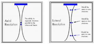
Using dark-field illumination with angles of illumination that exceed the numerical aperture, combined with Fourier pytchography, allows for a larger total NA. Having a larger NA increases lateral resolution. Lateral resolution is a subcategory of spatial resolution, and describes the ability to distinguish two points located perpendicular to the direction of the incoming beam of light. Lateral resolution is dependent on depth of field, with shallower depths having improved lateral resolution. This method also requires a low-magnification objective with a large field of view, helping to increase axial resolution, or the ability to distinguish two points parallel to the incoming beam of light. Together, the improved axial resolution and lateral resolution (comparison shown in the image below) contributes to high-resolution 3D intensity and phase images.
In order to recover 3D intensity and phase images from the captured data and remove aberrations that would cause blurred or distorted images, L. Tian and L. Waller use an algorithm called multislicing, which models the sample as a series of thin slices. The model also uses an iterative reconstruction routine that uses the light field measurements as an initial guess, and through a series of computations is able to eliminates out of focus blur and recover the images.
Overall, this paper proposes an effective alternative to confocal microscopy and other forms of microscopy for 3D imaging by improving temporal resolution while maintaining high resolution. This alternative uses a programmable LED array with dark-field illumination that can quickly and efficiently scan through a series of illumination angles, producing light field measurements that can be incorporated into an iterative reconstruction model for the fast and high quality production of 3D intensity and phase images.
References:
L. Tian and L. Waller . “3D Intensity and Phase Imaging from Light Field Measurements in an LED Array Microscope.” Department of Electrical Engineering and Computer Sciences, University of California, Berkeley. Optica 2. 104-111(2015).
D. Murphy, R. Oldfield, S. Schwartz, and M. Davidson. “Introduction to Phase Contrast Microscopy.” MicroscopyU.
W. Chambers, T. Fellers, and M. Davidson. “Darkfield Illumination.” MicrocopyU.
https://sites.google.com/site/ektasphysicseportfolio/axial-lateral-resolution