
Our most recent project involves imaging small, multi cellular aquatic organisms called hydra. This organism is most notably known for its lack of aging since it is comprised mostly of regenerative stem cells. Our hydra are provided by a lab in Swarthmore’s biology department. We were given vials containing about 10 fixed regular and ‘inverted’ hydra. The inverted hydra have been sliced open and turned inside out, providing another perspective of the hydra for imaging. We were also given a pipette, a small glass dish with a hollow in the center, and a stack of cover slips to rest over the hydra.
As we began our imaging procedure, we quickly ran into problems. For instance, because we are imaging with an inverted microscope, there is a hole in the microscope stage over the objective lens. The dish we were given for imaging the hydra was smaller than the hole in the stage, causing the dish to fall through and have no where to sit. To fix this, we acquired a larger dish, which did not have a center hollow.
We prepared hydra samples by pipetting one hydra onto the large dish and resting a cover slip on top. At first, this process appeared to work well. As imaging went on, however, we noticed that the body of the hydra seemed to expand and degrade. Below, we include the 1st and 137th images from one of our imaging datasets.
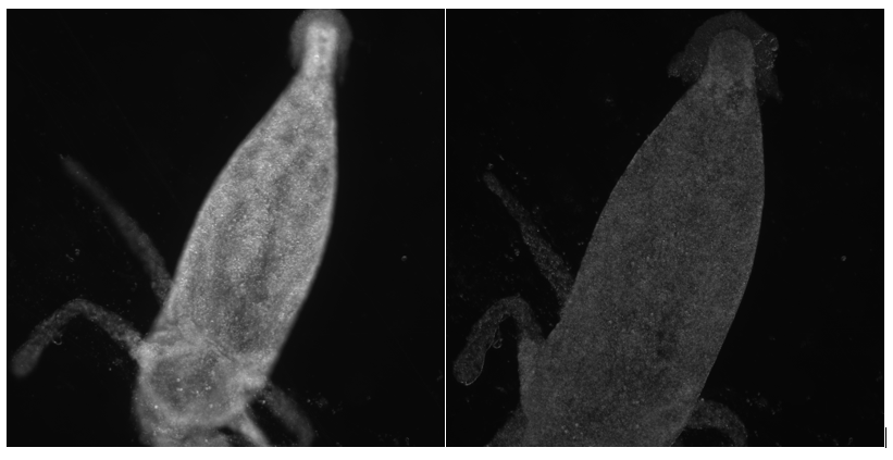
The hydra appears to have a different shape and density at the end of imaging than it did in the first image.
We brought our imaging results to our contact in the biology lab. We demonstrated our pipetting technique to show that the hydra were not being damaged during slide preparation. We mentioned that we had replaced the small dish with a larger one that had no center hollow because the small dish did not fit on the microscope stage.
The biologist explained that the dish hollow was an important feature that gave the hydra, a three-dimensional organism, space beneath the coverslip. When we applied the coverslip directly on top of our hydra, we were crushing it under the pressure of the cover slip. This caused the degradation we saw in our datasets.
He recommended building our own three-dimensional well for the hydra on a glass slide. By applying several layers of double sided tape on either side of the water droplet containing the hydra, we can raise the coverslip high enough that it does not crush the animal. This should prevent future sample degradation.