As we image and pre-process the hydra data, we have continued to encounter unexpected problems, leading us to make changes to our imaging procedure in order to best optimize the output of our data.
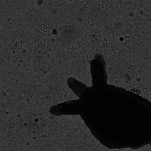
The first issue we encountered was the fact that the hydra were being flattened by the pressure of the cover slip while imaging was taking place. This caused the slow degradation of the hydra, causing major differences between image 0 and image 68 (see image below).
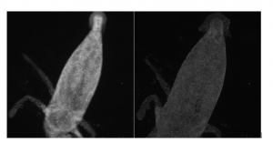
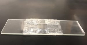
Differences between each of the 69 images in each stack, other than the change of position of the light source due to the each illuminated LED, are problematic and result in poor training data for the neural network. The lab group solved this problem by stacking up double-sided tape on the slides with the cover slip sitting on top, allowing for a 3-dimensional gap for the hydra to rest in during imaging.
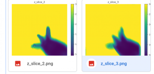
While the hydra itself are now better preserved during imaging, we have since come across additional imaging problems. We tried running our shift-add algorithm, which takes the low resolution stack of images and analyzes the image at different z-distances (depths), producing different three-dimensional “slices” of the image. However, we found that the resulting image slices were completely saturated with light, producing the yellow image below.
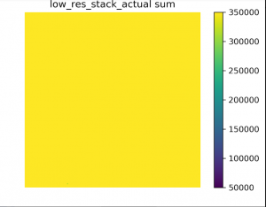
The lab group quickly realized that stray light from the lab room was saturating the images in too much light. In order to eliminate stray light, it is necessary to image either in a dark room or with a cover completely over the FP microscope. The lab group chose to use a large cardboard box as a cover for the microscope and camera. We took more data, ran the shift-add algorithm, and were successfully able to produce image slices of the hydra. Below is a look at an example of an original low resolution image of a hydra and a couple of its resulting slices from the shift-add algorithm.
The final problem, and so far one of the most difficult to solve, is the subtle movements of the water surrounding the hydra in the slide. These small movements of the water, caused by factors such as tiny shaking of the building, so small that they are nearly undetectable, cause the hydra to move ever so slightly in the flowing water. This results in a visual and problematic shift between image 0 and image 68 in a stack (example shown below), producing poor training data for the neural network.
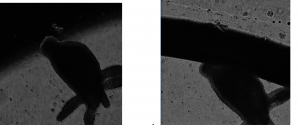
The ideal way to solve this problem is to use a specially designed optical table, which corrects for minute movements and shakes such as these. However, the lab currently is not equipped with an optical table, so alternative attempts at a solution have been made. Currently the lab group is using a tray with small dishes for imaging. We figure that with smaller dishes, the water has no place to move and expand, whereas with the taped slide method, there were empty pockets of space for the water to flow into. The potential drawback with this method is the tray is made of thicker plastic compared to the glass slide, which could possibly produce less clear images.